

A Subtle Scourge in the Bones
In the hills of northern Oman, beneath the collapsed stones of a 4,000-year-old tomb, archaeologists uncovered1 something that hadn’t been seen before in this part of the world: the unmistakable signs of leprosy.
It wasn’t a complete skeleton that told the story. It was fragments—broken jaws, scattered teeth, a porous nasal cavity eroded by years of silent bacterial assault. Pieced together from over 180,000 bone shards at the Umm an-Nar period site of Dahwa, the evidence pushed back the known boundary of Mycobacterium leprae’s reach. And in doing so, it also redefined what it means to diagnose disease in the archaeological record.
“When that damage fits key signs of leprosy, the diagnosis becomes very clear.”
So said bioarchaeologist Gwen Robbins Schug of the University of North Carolina at Greensboro, who led the team that identified lepromatous lesions in 12 individuals using micro-CT imaging—marking the earliest confirmed cases of leprosy outside of South Asia.

The Trade Route Nobody Wanted
The site of Dahwa wasn’t just a burial ground. It was a hub. Located along the copper-rich Al-Hajar Mountains and facing the Arabian Sea, it sat astride trade routes that once linked the Arabian Peninsula to the Indus Valley. Pottery fragments imported from South Asia—nearly three-quarters of the ceramic assemblage—testify to robust commercial ties. But alongside copper and crafted wares, it seems, another traveler moved: disease.
Leprosy is not easily transmitted. It requires extended, close contact. Most people have natural immunity. But in tightly packed settlements and among mobile traders moving between ecological zones and populations, it had the conditions it needed.
“The idea that sailors could have brought it on a crowded boat with them, or that merchants traveled from the Indus Valley toward Oman and spread it? Yeah, that makes a lot of sense,” said environmental historian Eric Strahorn, who was not involved in the study.
Seeing Beneath the Surface
Unlike many other cases of paleopathology, which rely on fully intact skeletons showing obvious signs of disease, the remains at Dahwa were fragmented and commingled. Traditional methods could only go so far. To push beyond them, the team used micro-computed tomography (micro-CT), scanning three preserved maxillae to look for the microstructural signatures of chronic infection.
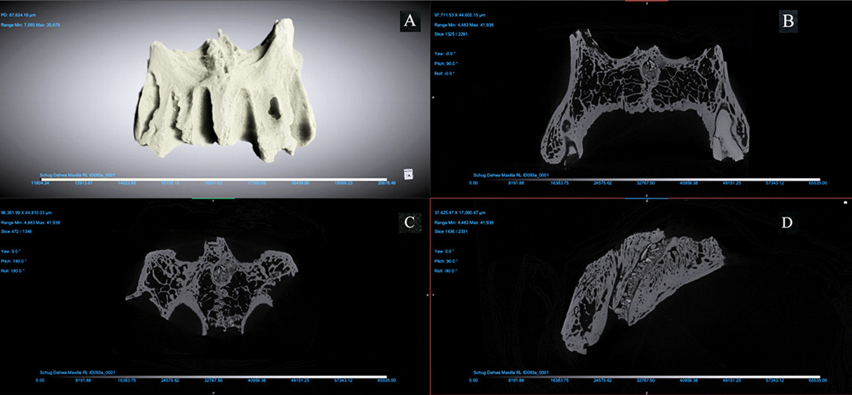
What they found matched what has long been considered pathognomonic for leprosy:
Resorption of the anterior nasal spine
Rounded erosion of the pyriform aperture
Bilateral alveolar bone loss in the upper jaw
Subperiosteal new bone formation on the palate
These changes were not random damage. They were patterned. And when compared to a control sample from modern anatomical collections, the distinctions became undeniable.
“Micro-CT lets us see what the eye can’t—subtle, patterned damage across multiple faces,” said Schug.
A New Tool for an Old Problem
Diagnosing leprosy has never been easy—not in antiquity, and not even today. Some modern patients present with atypical forms, delaying recognition until nerve damage and deformity have taken hold. In archaeology, the problem is compounded by time, taphonomy, and incomplete remains.
But Dahwa’s finds suggest a path forward.
“This paper adds to the limited number of studies evaluating rhinomaxillary changes using micro-CT in fragmentary and commingled assemblages,” the team wrote.
And beyond paleopathology, the implications stretch toward clinical medicine. In living patients, CT scans focused on the nasal aperture and ventral maxilla could supplement standard diagnostics—especially in ambiguous cases where microbial assays fall short.
Disease and the Human Condition
This isn’t just a story of pathogens. It’s a story of how humans adapt, organize, and endure. The people buried in Dahwa’s tombs were part of a larger economic and social landscape—one that spanned oceans and deserts, and that brought not just goods but biological entanglements.
As Robbins Schug puts it, “We’re learning how health, environment, and movement are deeply intertwined—even four millennia ago.”
The micro-CT data, stored and shared for future researchers, may help resolve other puzzles. And as climate change, migration, and zoonotic threats rise today, insights from ancient disease landscapes have renewed relevance.
Related Research
Robbins, G., et al. (2009). Ancient skeletal evidence for leprosy in India (2000 BC). PLoS ONE, 4(5):e5669. https://doi.org/10.1371/journal.pone.0005669
Köhler, K., et al. (2017). Possible cases of leprosy from the Late Copper Age (3780–3650 cal BC) in Hungary. PLoS ONE, 12(11):e0185966. https://doi.org/10.1371/journal.pone.0185966
Pfrengle, S., et al. (2021). Mycobacterium leprae diversity and population dynamics in Medieval Europe from novel ancient genomes. BMC Biology, 19:1–18. https://doi.org/10.1186/s12915-021-01120-2
Robbins Schug, G., Mahajan, S., Carter, S., Leach, A., Williams, K. D., Douglas, K. A., & Al-Jahwari, N. S. (2025). Lepromatous leprosy in Bronze Age Oman: micro-CT provides tools for paleopathology in fragmentary and commingled assemblages. Frontiers in Medicine, 12, 1521515. https://doi.org/10.3389/fmed.2025.1521515


